Systematic Position
Phylum
: Protozoa
Subphylum: Ciliophora
Class: Ciliata
Genus
: Paramecium
Species: caudatum
Occurrence
Paramecium caudatum is one of the
most common species of paramecium having worldwide distribution. It is found in freshwater
ponds, pools, ditches, streams, rivers, lakes, reservoirs, etc. It is usually
abundant in those waters which contain a great deal of decaying organic matter.
It thrives well in ponds or slowly running streams containing aquatic plants.
External Structure
- Size: Paramecium is a microscopic, elongated organism which is visible to the naked eye as a whitish or greyish spot. Its species vary in length from 80µ to 350µ. P. caudatum, the largest species, measures between 170µ and 290µ. The greatest diameter of the cylindrical body is about two-third of its entire length.
- Shape: Paramecium is slipper shaped. Its shape is constant and is asymmetrical. Because of its slipper-like shape it is sometimes called the slipper animalcule. The body is elongated blunt and rounded at the anterior end and somewhat pointed at the posterior end. In cross section, it is circular with greatest diameter behind the centre of body. The anterior half of body is slightly twisted. The body, is distinguished into an oral or ventral surface and an aboral or dorsal surface.
- Oral groove: Ventral surface of body bears a prominent, oblique and shallow depression, called oral groove. It originates from the middle of body and extends to the left side of anterior end. Posteriorly, the oral groove leads into a deeper conical vestibule which in turn communicates with a buccal cavity having a basal mouth or cytostome.
- Pellicle: External covering
of body is a living, clear, firm and elastic cuticular membrance, the pellicle. When observed under light
microscope, the pellicle appears to be a regular series of polygonal (or
hexagonal) depressions with their raised rims. A single cilium emerges out
from the middle of each polygonal space.
- Cilia: The
entire body surface is covered by numerous, tiny,
hair-like fine projections, called cilia. These measure 10-12µ in length one
cilium arises from the centre of each polygonal depression (circumciliary
space) of- pellicle. There are 10,000 to 14,000 cilia covering the whole-body
surface. These motile organelles are arranged in regular longitudinal
rows. They remain equally distributed though out body surface. Their
length is uniform throughout, except for a few longer cilia at the extreme
posterior end of the body, forming a caudal tuft, hence the
species name caudatum is given.

Internal Structure:
1. Cytoplasm: - Inside
pellicle,
the cytoplasm of body is clearly differentiated into two regions.
(a) Ectoplasm: The narrow,
peripheral, clear and dense region is called the ectoplasm. It consists of the
structure of the infraciliary system and the trichocysts.
(b) Endoplasm: The large,
central, granular and semi-fluid region is the endoplasm. It consists of
the usual cell components like mitochondria, Golgi bodies, ribosomes, reserve
food granules, etc. Prominent endoplasmic inclusions are nuclei, contractile
vacuoles and food vacuoles.
2. Infraciliary system: Immediately
beneath the pellicle the infraciliary system constituted by the basal
bodies and kinetodesmata is present.
(a) Basal
bodies: The base of each cilium is produced into a tube-like
structure, called basal body or kinetosome. The basal bodies are
self duplicating units and progenitors of new cilia. Each basal body is
either a centriole or its derivative.
(b) Kinetodesmata:
Associated
closely with basal bodies of cilia and lying in the ectoplasm is a system of
specialized striated fibrils, called kinetodesmal fibrils. A single
fibril or kinetodesmos arises from the kinetosome or basal body of each
cilium and runs anteriorly somewhat tapering along the, course.' It joins its
counterparts from the posterior kinetosomes, forming a bundle of overlapping
longitudinal fibrils, called kinetodesma (pleural, kinetodesmata). The
number of fibrils in each kinetodesma remains constant (5) because the
individual fibrils do not run anteriorly farther than 5 basal bodies. It
has been suggested that fibrils coordinate ciliary beat and movement, but the
evidence is very conflicting.
The kinetosomes of a longitudinal
row. plus their
kinetodesmata constitute a structural unit, called the kinety. A kinety system is apparently characteristic of all ciliates.
3. Trichocysts: Trichocysts
are peculiar rod like or oval organelles present throughout the ectoplasm
alternating with basal bodies and oriented at right angles to the body surface.
These are very small in size, measuring about 4µ in length.
Each trichocyst consists of an elongated shaft and a terminal pointed tip,
called the spike or barb, covered by a cap. The
matrix of shaft consists of a dense mass of a fibrous protein, called trichinin.
Function of trichocysts is not well known. It is believed
that these discharges and anchor the animal to a firm substratum when it feeds
upon bacteria.
Others believe that these are organelles of defence.
Discharge of trichocysts is triggered by mechanical,
chemical or electrical stimulation. It occurs. in a span of a few milliseconds. When fully
discharged, the shaft becomes a long cross-striated rod and measures about 40µ in length.
4. Nucleus: Paramecium
is
heterokaryotic
as
it
possesses two types of nuclei. In P. caudatum, there is a large macronucleus and a small micronucleus. Besides the
macronucleus, two micronuclei are present in P. aurelia and many in P.
multimicronucleatum.
(a) Macronucleus:
The
macronuclens
is
roughly kidney-shaped and with inconspicuous nuclear membrane. It is polyploid
and possesses many nucleoli and much more chromatin material (DNA). Macronucleus
is the somatic or vegetative nucleus and controls the day-to-day metabolic
activities of the cell.
(b) Micronucleus:
The
micronucleus
is
present
in a depression on the surface of the macronucleus. It is usually spherical,
with a nuclear membrane and with diploid number of chromosomes.
5. Contractile
apparatus: In Paramecium, there are two contractile
vacuoles, occupying somewhat fixed positions in endoplasm. One
vacuole lies near each end of body, close to the dorsal surface. Each of them
is surrounded by a circlet of 6 to 10 long, narrow, spindle-shaped radial canals extending far
into cytoplasm. Each contractile vacuole opens to outside through a permanent
pore in pellicle of dorsal side of body. The two contractile vacuoles do not
lose their identity when water is expelled.
Each radial canal consists of terminal part, ampulla and
injector canal. The radial canal collects liquid from large part of body and
pour it into vacuole. When vacuole attain maximum size, it collapses
discharging its contents to the outside. It is osmoregulatory and excretory in
nature.
6. Food
vacuoles: Numerous non-contractile food vacuoles, or gastrioles are present
moving with the streaming endoplasm (cyclosis). They differ in shape and size
according to the nature of ingested food particles, but mostly they are rounded
in form.
7. Oral
apparatus: In Paramecium, oral groove
leads ventrally and posteriorly as a tubular structure, called vestibule. It leads
directly into a wide tubular passage, the buccal cavity. In its turn,
it opens into a narrow gullet or cytopharynx through a
narrow aperture, the cytostome. The
cytopharynx, at its proximal end, forms a food vacuole.
8. Cytopyge:
Near
posterior end of body, a little behind cytostome a small portion of ectoplasm
and pellicle is somewhat weak. Here, at the time of egestion, a minute
aperture called cell anus, cytopyge or cytoproct, is visible. It
is, however, difficult to say whether it is a permanent opening with tightly closed
lips or a temporary opening formed at the time of egestion.
Reproduction:-
Parameium reproduces
asexually by transverse binary fission and also undergoes several kinds of
nuclear reorganizations, such as conjugation, endomixis, autogamy, etc. Under
certain conditions of food and temperature, it undergoes encystment.
1. Transverse binary fission
During favourable conditions, Paramecium commonly reproduces by
transverse or horizontal binary fission. During it paramecium stops feeding and its oral groove and buccal
structures begin to disappear. While this is happening, the micronucleus starts dividing by the complicated
process of mitosis, into two daughter micronuclei. The daughter micronuclei
then separate. Simultaneously, the macronucleus divides amitotically by
simply becoming -elongated and constricted in the middle. Two oral grooves now begin
to form, one in the anterior half and the other in the
posterior half. Two original contractile vacuoles remain, one in each half of the
dividing parent individual. Two new contractile vacuoles are later formed. Two
new buccal structures also appear. In the meantime, a constriction furrow
appears near the middle of body. It deepens and ultimately the cytoplasm is
completely divided, resulting into two daughter paramecia. Of the two
daughter paramecia, the anterior one is called proter and the posterior,
opisthe. These grow to full size and divide again by
fission.

P. caudatum divides 2-3 times in. a day by binary
fission. The process is completed in about 30 minutes, though separation of
daughter paramecia takes about one hour or more. The term clone refers
to all the individuals that are produced asexually from one parent paramecium.
All the members of a clone are genetically alike.
2. Conjugation
Paramecium undergoes a sexual phenomenon, which is called conjugation.
It is frequently referred to as sexual reproduction, but it is simply a temporary
union of two individuals of one and the same species for the purpose of
exchanging a part of their micronuclear material. This remarkable process in Paramecium
occurs frequently between binary fissions and' is necessary for the
continued vitality of the species.
1. Process of conjugation: In conjugation,
two individuals or preconjugants, from two different mating types, come
in contact ventrally and unite by their oral grooves. They stop feeding and
their buccal structures disappear. The pellicle and ectoplasm degenerate at the
point of contact and a protoplasmic bridge is formed between the two
individuals, which are now called the conjugants. While so united, the conjugating
pair continues to swim actively and a sequence of complicated nuclear changes
takes place in each animal.
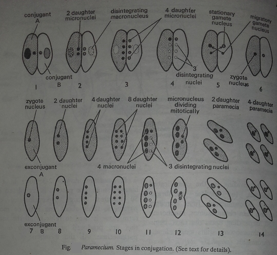
The vegetative macronucleus simply breaks
up into fragments and disappears. The
diploid micronucleus
of each conjugant first grown in size and then
divide by meiosis. Thus, 4 haploid daughter micronuclei are produced of
which 3 degenerate and disappear in each conjugant, while the remaining one
divides by mitosis forming 2 unequal pronuclei or gamete nuclei.
The
smaller one is the active migratory gamete nucleus and the bigger
one is the passive stationary gamete nucleus. The migratory
nucleus of one conjugant then passes through the protoplasmic bridge into the
other individual and fuses with its stationary nucleus, forming a single
diploid zygote
nucleus or
synkaryon.
The
complete fusion of two nuclei from two different individuals forming a zygote
nucleus is termed amphimixis.
The two pairing paramecia, after a union of about 12 to 48
hours, separate and are now called exconjugants. In each
exconjugant, the zygote nucleus divides by mitosis three times in rapid succession
producing 8 nuclei, of which 4 enlarge to become macronuclei and other 4
become micronuclei.
Three
micronuclei disintegrate and disappear, while the remaining micronucleus
divides, with binary fission of exconjugant. Thus, from each exconjugant two daughter
paramecia are obtained, each containing two macronuclei and one mincronuclei. The
micronucleus again divides with the division of each daughter paramecium,
forming two
individuals
each containing one macronucleus and one micronucleus. Thus, each conjugant
produces four daughter individuals at the end of conjugation.
2. Factors and
conditions of conjugation. Conjugation is very complex physiologically.
The factors and conditions governing conjugation are several and these may also
vary with the species.
1. Conjugation
does not occur under favourable living conditions. Starvation or
shortage of food and a
particular bacterial diet or certain chemicals are said to
induce conjugation
in some special.
2. A
certain range of, light and temperature is said to be essential, for
conjugation.
3. In
P. caudatum,
conjugation
usually starts early in morning and is continued till
afternoon.
4. The conjugating
individuals are usually smaller in size than the normal individuals.
5. Conjugation
never takes place among the members of a "pure line", that is among the descendants of a single individual. It occurs only between individuals
belonging to two different mating types. Thus, a sort of
physiologically sexual differentiation exists in Paramecium.
3. Significance
of conjugation: The significances of conjugation are:
(a) Rejuvenation. If binary
fission continues repeatedly for several generations, the Paramecium loses its
vigour and enters upon a period of depressed physiological efficiency and
senescence. The individual ceases to multiply, reduces in size, degenerates in
organization and eventually dies off.
b) Nuclear reorganization.
During
conjugation, the nuclear apparatus is reorganized and a readjustment occurs
between it and the
cytoplasm,
Probably the macronucleus loses its potentialities in performing its manifold
metabolic activities. Its replacement by a new macronucleus brings renewed
vigour and vitality to accelerate the metabolic activities. '
C) Hereditary variation. During asexual
reproduction by fission, the hereditary material of the parent passes unchanged on to the progeny, so that all the
descendants of one Paramecium have the same
inheritance. The periodic occurrence of conjugation, however, ensures inherited variation. It brings
about, the blending of two lines of ancestry just as bisexual reproduction
does.
4. Genetic consequences of conjugation: If conjugation
takes place between two paramecia, one homozygous for a dominant
gene (AA)
and
the other homozygous for its recessive gene (aa), the first generation would be
heterozygous (Aa). If the two conjugants are already heterozygous (Aa), then the
resulting progeny would be either homozygous or heterozygous, depending upon
which gene gets eliminated at the stage of disintegration of three micronuclei
in each conjugation.