Connective tissue
It is mesodermal in origin. The components of connective tissue are matrix, fibres and cells. Matrix is the ground substance which contains different organic and inorganic substances. It may be gelatinous or amorphous or transparent. Intercellular spaces are filled with large amount of extracellular material of protein fibers lying in amorphous transparent ground substance Matrix. Fibers are secreted by cells. Matrix is fiber free in vascular or fluid connective tissue while matrix is densely mineralized in bone.
Functions of connective tissue:
*It binds different structure muscles with skin, Bone with bone, bone with muscles etc.
*It forms protective sheath around delicate organs like kidney, spleen, testis, heart etc.
*Skeletal tissue provides support and forms framework of body.
*Adipose tissue stores fat.
* Fluid connective tissue is responsible for transport.
* The phagocytes are responsible for protection.
*Builds immunity by producing antibodies.
Types of connective tissue
Connective tissues are categorized on the basis of nature of matrix. Three types of connective tissue are found which are Connective tissue proper, Supportive connective tissue and vascular or fluid connective tissue.
Connective tissue proper: The matrix is soft, less rigid with varying degree of toughness. It binds different organs and parts of various organs together. On the basis of amount of fiber, it may be loose connective tissue and dense connective tissue.
Loose connective tissue: Less fibre are present and are loosely woven. The cells are widely distributed in matrix. It is of two types; areolar tissue and adipose tissue.
Areolar tissue:
It consists of homogenous, semi-fluid, transparent and gelatinous matrix. The matrix is mixture of glycoprotein, mucin, hyaluronic acid and chondroitin sulphate. In matrix various kinds of cells and fibres are present.
 |
| Areolar tissue |
Types of cells:
Four types of cells are found which are;
Fibroblasts are spindle shaped flat cell with Oval nucleus and protoplasmic processes. They produce fibers.
Macrophages are large amoeboid cell with kidney shaped nucleus. They engulf bacteria, Ingest damaged tissue.
Mast cells are large Oval cell with granular cytoplasm. They produce Heparin (Anticoagulant), Histamine (inflammatory) and serotonin (Vasodilator).
Plasma cells are Small round cells and produce antibodies for self-defense.
 |
| Types of cells |
Types of fibers:
The fibers may be collagen fibre, elastic fibre or reticulin fibre.
Collagen fibers also called as white fibre are made-up of protein collagen. They always occur in bundle and are wavy. They are thick, flexible but inelastic with great tensile strength. When boiled in water collagen changes into gelatin.
Elastic fibers are also called as yellow fibers. They are formed of protein elastin. They are long, straight, branched and found single. They are thin and elastic in nature.
Reticulin fibers are formed of protein reticulin. They are fine, short, thread like and occur singly. They are delicate and are found in network. They are regarded as immature collagen fibres.
 |
| Types of fibres |
Adipose tissue: It is modified form of areolar tissue. It is specialized loose connective tissue in which the fibroblasts are modified for fat storage. The fat storing cells are called as adipocytes Or fat cell. They are large, spherical or Oval shaped almost filled with fat droplets. Adipocyte with many small fat droplets is called brown adipocyte while with asingle large fat droplets is called White Adipocyte.
Adipose tissue also contains fibroblast, macrophages, Collagen and elastin fibers. Adipose tissue is found beneath the skin, around kidney, heart, eyeballs etc. Adipose tissue synthesizes, stores and metabolizes fat. Adipose tissue is a poor conductor of heat. It reduces heat loss through skin. It acts as shock absorber around heart, kidney, eyeballs etc. Brown adipocytes give more energy and stores more energy in the form of glycogen.
 |
| Adipose tissue |
Prominent adipose tissue sites are subcutaneous fat, Blubber of whales, Hump of camel, in frog adipose tissue forms the fat bodies.
Dense connective tissue: Connective tissue proper with more fibers is called as dense connective tissue. More fibers are present in comparison to matrix and cells. Fibers may be regularly arranged or irregularly arranged. It is of three types white fibrous tissue, yellow fibrous tissue and reticular tissue.
White fibrous tissue: It is found in tendons, some ligaments, sclera of eye, Cornea of eye, kidney capsule etc. and is also present in perichondrium of cartilage and periosteum of bone.
It is tough, shiny and composed of numerous highly organized bundle of collagen fibers. Collagen fibers are tightly packed together running parallel to each other. Rows of Fibroblasts run alongside the bundle of fibers. Each bundle bounds to it’s neighbor by areolar tissue. It gives tensile strength. It is present in the joints of skull bone and forms almost immovable joints due to the presence of inelastic collagen fibres.
 |
| White fibrous tissue |
Tendons: It composed of almost entirely of parallel collagen fibers aggregated to form bundle. Fibers are bound together by matrix; tendomucoid. Only fibrocytes are present along the bundle of fibers. It lies inside fibroclastic tissue (sheath). It gives strong, flexible/pliable but inextensible strength. It joins skeletal muscle to bone. The rupture of tendon is called as strain.
Sheath is also white fibrous tissue which forms protective thin layer or covering around different organs.eg. perichondrium, periosteum, sclera/ cornea of eye etc.
Ligaments ( Yellow fibrous tissue): have loose network of irregularly arranged branched yellow fibers and some collagen fibers. It is yellowish due to elastic fibers. Fibroblasts are present. It is elastic in nature. It is found in vocal cords, wall of artery, Bronchioles etc. It joins bone with bone. Sprain is the stretch of ligament.
 |
| Tendon and Ligament |
Reticular connective tissue: It consists of network of reticulin fibres. It is found in spleen, liver and lymph. It contains phagocytes and forms defense system of body and protects body from infection. Reticular cells with protoplasmic processes are present. The matrix is fluidy.
Skeletal or
supportive connective tissue
It is with mineralized matrix. It is of two types; cartilage and bone.
Hyaline cartilage: It is more abundant cartilage. It adds articular surface at joints (ends of the bones). It forms coastal cartilage of ribs. It is found in nose, larynx, trachea, bronchus etc. Most of the embryonic skeleton is of hyaline cartilage. The matrix is glass like semi transparent and fine collagen fibers are present with fiber free appearance. It is slightly elastic and compressible.
Elastic cartilage: Matrix is Semi-opaque with network of yellow fibers. It is highly elastic and flexible. It recovers the shape quickly. It is found in external ear, epiglottis and Eustachian tube. It is also called as yellow cartilage.
Fibrous cartilage: It is also called as white fibrous cartilage. The matrix has bundle of densely packed collagen fibres. It provides greater strength and little flexibility. It acts as shock absorber. It is found in between adjacent vertebra as intervertebral disc and in pubic symphysis of pelvic girdle.
 |
| Types of cartilages |
Calcified cartilage is cartilage in which matrix contains salts of calcium.
It is hardest tissue and forms endoskeleton. Its matrix is solid and calcified. It is made-up of cells embedded in matrix. The matrix consists of organic and inorganic substances and protein ossein (formed by osteoblasts). About 70% Inorganic substances are found in matrix and consists of Phosphates, Sulfates, Carbonates of calcium and magnesium. The main inorganic substance is hydroxyapatite [Ca10(PO4)6(OH)2]. All these makes bone hard. Along with age these Inorganic matter decreases and bones become brittle. Long bones are hollow Inside and the cavity is called marrow cavity which is filled with bone marrow. Bone marrow is of two types; red marrow and yellow marrow. Red marrow forms erythrocytes and granular leucocytes. The yellow marrow is for storage of fat. The ends of long bone are called as epiphysis While shaft or the main part is called is diphysis.
Structure of bone: Each bone is enclosed by a layer of white fibrous tissue called periosteum. The matrix is arranged in concentric circles called as lamellae. In between lamellae bone cells called osteoblasts or osteocytes are present in fluid filled cavities called as lacunae. Osteoblasts are active bone cell while osteocytes are inactive bone cell. Each lacunae has fine cytoplasmic extensions called as canaliculi which passes through lamella and make connections with other lacunae. In compact bones, these lamellae are present around a canal called as Haversian canal. Haversion Canal, Concentric lamellae and lacunae with Canaliculi form Haversian canal system which is a special feature of bone. An artery and vein Pass through haversian Canal and through Canaliculi pass into lamellae. Haversian canal system is also called as Osteon. The horizontal canals which connect different Haversian canal are called as Volkmann’s canal.
 |
| Structure of bone |
Types of bone: Bones are of two main types; compact bone and spongy bone.
Compact bone: The matrix is solid, hard and dense. They form shaft of long bones (Diphysis). They are filled with yellow marrow which stores fat. The Haversian Canal system is present and is organised. There is no gap between lamellae hence they are compact.
Spongy bone: Matrix has network of thin interconnected trabaculae and forms the ends of long bones( Epiphysis). They are filled with red marrow which forms RBC. They are devoid of organized Haversian canal system. There is presence of gap between Lamella hence they are spongy.
*Osteoblasts are active bone cell.
*Osteocytes are inactive bone sale.
*Osteoclasts are cells which absorbs calcified matrix from bone. They are also called as Bone Cutter cell.
It is a special type of connective tissue in which matrix is fluidy and fiber free. The matrix is not secreted by cells. The cells have no power of division. It is responsible for internal transport and play role in defense mechanism.
The fluid present outside cell is called as extracellular fluid. It includes blood, lymph, coelomic fluid, Pericardial fluid, pleural fluid, interstitial fluid, cerebrospinal fluid, aqueous humour, endolymph, perilymph etc. Total extracellular fluid is about 15 ltrs. It contains about 45% water of body.
The vascular tissue is of two types; blood and lymph.
Blood: It is opaque, viscous, heavier than water and slightly alkaline with 7.4 Ph. The oxygenated blood is bright red colored while deoxygenated blood is purple. The study of blood is called as haematology. About 5-6 ltrs of blood is found in male while 4-5 ltrs in female. Main buffer is sodium bicarbonate while Ph is maintained by sodium bicarbonate and Carbonic acid.
It consists of Plasma and formed elements.
 |
| Composition of blood |
Plasma: About 55% , pale yellow, nonliving and consist of water(90-92%), inorganic salts(1%), plasma protein(7%) and organic substances(1-2%).
Plasma contains nutrients like glucose, amino acids etc,excretory products, hormones, vitamins, gases, antibodies, bacteria etc.
Plasma proteins are albumin globulin and fibrinogene/ prothrombin.
Albumin is smallest, most abundant and most important. It maintains osmotic pressure of blood and retains water. If albumin amount decreases, Water retention capacity decreases. Thus, water passes out from blood into tissue which results into odema causing swelling of hands and leg. It also regulates normal blood pressure and osmoregulation.
Globulin: Is responsible for defense mechanism. It is mostly produced in liver.
It may be; a-globulin, b-globulin,y-globulin and prothrombin. Y-globulin acts as antibodies and give immunity. Prothrombin helps in blood clotting.
Fibrinogen Is responsible for all blood coagulation. It forms insoluble fibrin that forms blood clot.
Functions of plasma: It is responsible for transport, immunity, regulation of osmotic pressure, temperature regulation, Clotting of blood, transfer heat etc.
Formed elements or blood corpuscles:
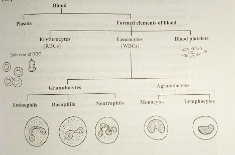
The blood cells are called formed elements or blood corpuscles as they don’t have all components of a typical cell. They are of three types; erythrocytes, leucocytes and platelets.
Origin of blood corpuscles:
*Embryo up to nine months in Yolk sac.
*Foetus in liver, spleen,Thymus, lymph nodes, bone marrow.
*Adult in bone marrow (Myeloid tissue) and Lymphoid tissue(spleen and lymph nodes).
Erythrocytes: Also called as red blood cells or corpuscles . A red colored blood cell. In human; It is small, biconcave and enucleated. They are without mitochondria, centrosome, Golgi body, ribosomes etc. Due to which more haemoglobin can be stored. Due to the absence of mitochondria, it becomes more efficient to carry oxygen. It respires anaerobically and it helps to carry more oxygen. It needs less energy. Its life span is about 120 days. The process of formation of RBC is called as erythropoiesis which takes place in bone marrow, myeloid tissue. Erythropietin is a hormone produced by kidney which stimulates erythropoiesis. Vitamin B12 and folic acid are required for maturation of red blood cell. Iron containing pigment haemoglobin is present in red blood cells which carries oxygen. Haemoglobin has iron part haeme and protein part globin. About 4-5 million RBCs are present per cubic cc of blood. Normal haemoglobin level is 12-14 gram per 100 milliliter of blood in adult while 22 -23 gram per 100ml of blood in newly born child. Destruction of RBC takes place in liver and spleen. Spleen is called as graveyard of RBCs. About 2-10 million RBCs are destroyed and formed in a second.
*Anemia is fall in RBC.
*Polycythemia is rise in RBC.
Haemoglobin iron containing respiratory pigment found in RBC and gives red colour while other respiratory pigments are;
Haemocyanin copper containing pigment found in plasma and gives blue colour. It is found in arthropods and molluscs.
Chlorocruorin iron containing pigment found in plasma and gives green color. It is found in some annelids.
Haemoerythrine iron containing pigment found in corpuscles and gives red colour. It is found in some annelids.
In Mammals largest RBC is found in elephant while smallest in musk deer. In vertebrates; largest RBC are found in amphibians; Amphiuma(Salamander).
In Camel and Llama Oval and nucleated RBCs
are present.
Cyanosis is blue colored skin due to less
hemoglobin.
Leucocytes are of two types, agranulocytes and granulocytes.
Granulocytes: Are with enzyme rich granules of lysosomes.
Agranulocytes: Granules are not present.
Granulocytes: About 72% granulocytes are present. They are of three types; Eosinophils, Basophils and Neutrophils.
Eosionophils also called as acidophils. They are stained by acidic dyes like eosin. Their nucleus is bilobed. They produce antihistamine. They are about (1 to 3)%.
Basophils are stained by basic dyes like methyl blue. Their nucleus is S- shaped. They produce histamine, heparin and Serotonin. They are about 1%.
Neutrophils are stained by neutral dyes. They have multilobed nucleus and are phagocytic in nature and engulf bacteria. They are about( 68 -70%.
Agranulocytes: About 28%. They are of two types, monocytes and lymphocytes.
Monocytes are with large bean shaped nucleus. They engulf bacteria and are Phagocytic in nature. They are about 4%.
Lymphocytes are with large Oval nucleus. They produce antibodies. They are also phagocytic in nature. They are of two types, T-lymphocytes and B-lymphocytes. T-lymphocytes are produced in thymus while B-lymphocytes are produced in bone marrow.They are about 24%.
*Leukemia or blood cancer is uncontrolled
production of WBC.
*High WBC is called as Leukocytosis.
*Low WBC is called as Leucopenia.
They are irregularly shaped bodies smaller than RBC. They are without nucleus. They are fragments of large bone marrow cells called as megakaryocytes. About 2, 50,000 per cubic mm of blood. Their life span is about seven days. They are responsible for coagulation of blood. Their site of destruction is liver and spleen. Thrombocytosis is Rise in number of thrombocytes while Thrombopenia is decrease in number of thrombocytes.